// Viral RNA showcase — not a locked-benchmark cell
Druggability extends beyond riboswitches to pre-organized viral RNA elements — this is the evidence. The binding site here is mapped by sequence alignment from a different PDB entry (3TZR, a 2-strand crystallization construct carrying the ligand) onto the folded target (1P5M, the apo NMR structure) — not a same-structure co-crystal like the seven riboswitch targets on the locked benchmark. A whole-structure 3D overlay (like the FMN apo→holo demo) isn’t valid here — 3TZR splits the hairpin into two separate strands for crystallization, a different topology from the continuous predicted fold. The ligand toggle below instead uses a local superposition, fit only on the residues that align unambiguously between the two structures (fit RMSD 5.3 Å across 30 residues) — clearly labelled as approximate, not a validated co-crystal placement. The mapped binding-site residues are also shown in the sequence block below either way.
RNA pocket discovery
HCV IRES domain IIa · 1P5M
Bound by a small-molecule HCV IRES domain IIa binder, chromeno-imidazole class (PDB ligand code SS0; verified via the RCSB chemical component dictionary) (PDB ligand SS0). 55 nt RNA target.
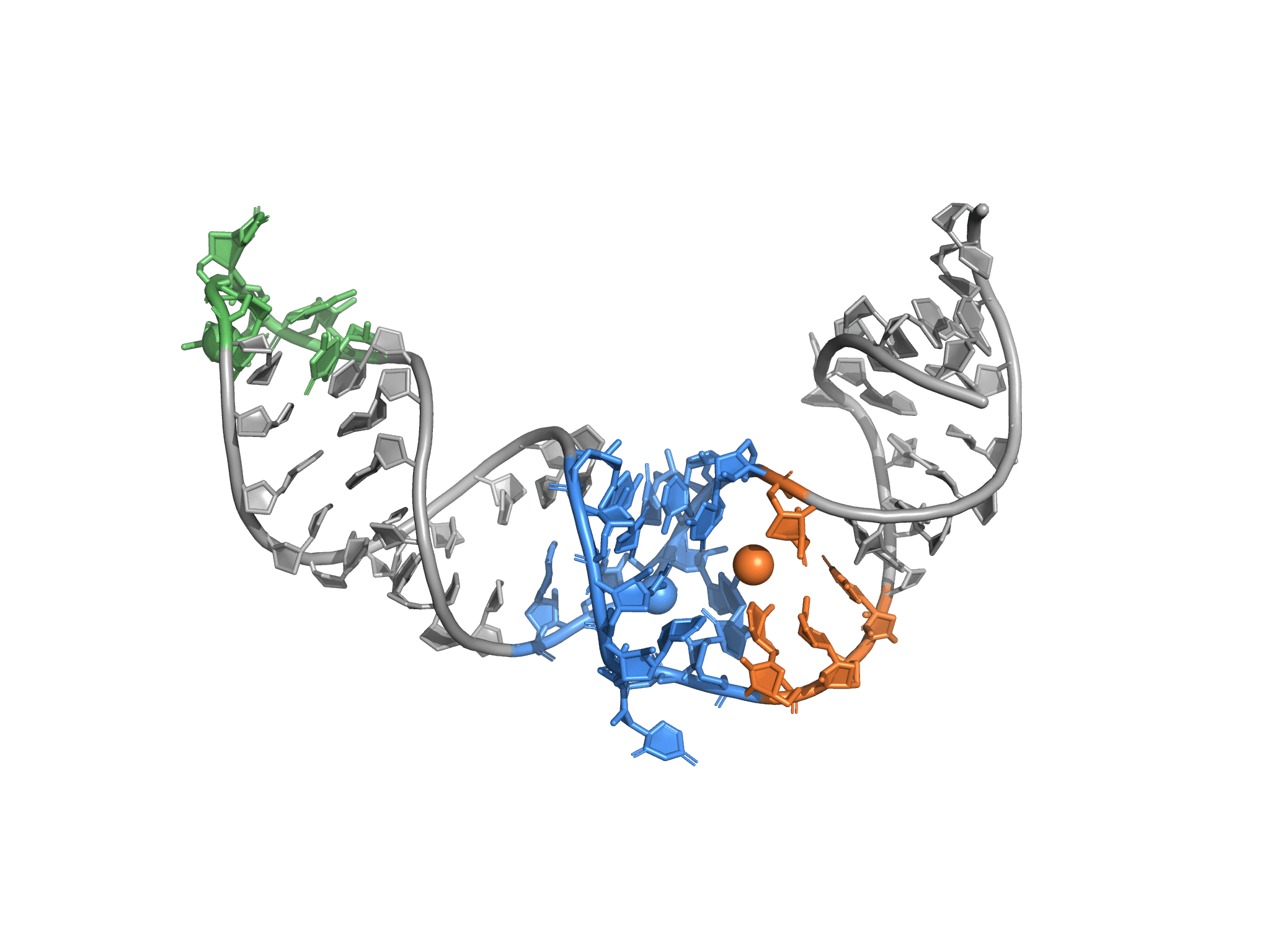
Top-3 candidate pockets
ranked by persistence × binding-residue stabilityCartoon backbone of the predicted reference frame. Top-3 pocket residues highlighted as licorice; centroid spheres mark each cluster's geometric centre across the ensemble. Hover a card to isolate that pocket. Toggle the ligand overlay — approximate, not a co-crystal placement: mapped from a different crystallization construct via local superposition on the sequence-aligned region only, not a whole-structure fit. See the caveat below for the fit quality.
Sequence with pocket residues highlighted
Methods summary
v0.2 detects cavities on the predicted 3D structure using RNA-tuned fpocket parameters (consistent with the published fpocketR approach, Veenbaas et al. PNAS 2025), samples a 5-frame ANM conformational ensemble, and ranks pockets by structural persistence weighted by binding-residue stability (score = persistence × n_residues_intersected). The cross-frame geometric ranker is the load-bearing contribution: on a 7-target cleft-binder benchmark, RNA-tuned detection alone recovers 0 of 7 at strict@1; the ensemble + ranker lifts recovery to 3 of 7 strict@1 and 6 of 7 near@1. Druggability assessment itself is left to your medicinal-chemistry workflow; we provide the geometric metadata and conformational stability metrics as inputs to it. Computational predictions only — experimental validation is required before use in drug development.
Read full methodology →Binding-mode caveat
Pipeline detects cleft-shaped binding pockets. Groove-binding modes and shallow surface-deformation binding may be missed. Contact us if your target's binding mode is groove-mediated.
Computational predictions only. Experimental validation required before drug-development use.
// How to read this result
- This is a cross-referenced showcase, not a ranking benchmark. The locked rank-1 benchmark (seven riboswitch targets, same-structure co-crystals) lives on the methodology page.
- The structures. Folded target: PDB 1P5M (apo NMR, chain A, 55 nt). Ligand site source: PDB 3TZR (2-strand crystallization construct carrying ligand SS0, a chromeno-imidazole HCV IRES IIa binder). The binding-site residues are mapped onto 1P5M by sequence alignment (longest-common-substring) — the same method used for the locked benchmark’s peptide- and literature-derived sites.
- The 3D ligand overlay is an approximation, not a co-crystal placement. 3TZR’s two-strand construct has a different global topology from the continuous predicted fold, so a whole-structure superposition isn’t valid. The toggle instead uses a local Kabsch fit restricted to the 30 residues that align unambiguously between the two structures (fit RMSD 5.3 Å) — stated explicitly every place the overlay appears. As an independent sanity check, not a hidden assumption: the placed ligand’s nearest atom lands 3.3 Å from the pipeline’s independently-detected rank-1 pocket, consistent with the strict@1 result below.
- Pipeline unchanged. Same single-sequence RhoFold + 5-frame ANM ensemble + geometric ranker as every other worked example — no target-specific tuning.